1. The serpentarium jewels
Despite the widespread idea that snakes are common animals in the wild and rural areas, the truth is that they are elusive, cryptic, and uncommon reptiles, even more so in the tropics. Finding snakes in tropical ecosystems is normally a hard task. Most snakes live in secretive, isolated, and difficult-to-access habitats (e.g., hyperhumid Chocoan rainforest, Amazonian rainforest). Because of armed conflict in Colombia, some parts of the country have been historically inaccessible or highly risky for researchers [1,2].
Achieving a robust sampling of tropical snakes is always a major challenge, and even more if the goal is to capture venomous species to establish a large-scale snake captivity program for antivenom production. Historically, experienced researchers have employed intensive survey designs in defined areas in tropical habitats, demonstrating that even devoting a tremendous sampling effort is not sufficient to achieve a completely representative sample of the local snake community [3–5].
So, the acquisition of live snakes for antivenom production, attempting to establish a geographically representative sampling that covers all snakes that are medically important, demands challenges such as: (1) building a strong collaborative partner network that includes researchers, governmental, and non-governmental allies; (2) investing much effort performing intensive snake sampling; and (3) being patient and having a high tolerance for frustration due the elusive and cryptic nature of snakes. This is why all snakes for antivenom production collected across the decades and housed in the Colombian National Health Institute (INS, Spanish acronym) are jewels that deserve to be extremely well-cared for and carefully maintained. This chapter summarizes the history of learned lessons about the venomous snakes housed in the serpentarium of the INS, as well as the most remarkable information about these serpentarium jewels.
1.1. Taxonomic richness and snake representativeness
Historically, the serpentarium of the INS has housed at least 608 snake specimens, according to the clinical records available from 1990 to 2021. However, this number is far from representative of the true history of the serpentarium because during the period 1965-1989 when it was located at the municipality of Armero (Tolima department), around 1,000 snake specimens were kept (com pers. Juan Manuel Renjifo, see Chapter 7). However, records of these specimens are scarce because standardized clinical records were not established until the year 2000 (Table 1).
During the early stage of the serpentarium in Armero, all housed snakes were represented by three species: Crotalus durissus (cascabel),Bothrops asper (lancehead pitviper),and B. atrox (mapanare). After the relocation of Armero’s serpentarium (see later) over time, the snake abundance, and species richness of the serpentarium grew gradually, adding occasionally pit vipers like Bothriechis schlegelii (eyelash pit viper), Lachesis muta (Amazon bushmaster), but maintaining as core species for antivenom production Crotalus durissus,Bothrops asper,and B. atrox. From 2012 to the present, a noticeable increase in abundance and species richness of medically important Colombian snakes was achieved (Table 1), incorporating for the first time as permanent species in the collection coralsnakes (Micrurus spp.) in the snake production population. These achievements strengthened national antivenom production and allowed the establishment of a large-scale intensive snake captivity program for coralsnake antivenom production [6].
Table 1. Number of venomous snakes housed historically, the serpentarium of the National Health Institute of Colombia.
|
Period
|
Family Elapidae
(Coral snakes)
|
Family Viperidae
(Pitviper snakes)
|
Total specimens
|
|
1965-1989
|
0
|
~1,000
|
~1,000
|
|
1990-2011
|
0
|
49
|
49
|
|
2012-2021
|
181
|
378
|
559
|
|
Total
|
181
|
427
|
608
|
Across the last decade, the serpentarium has maintained seven of the eight genera of snakes medically important in the country: pitvipers (Bothriechis, Bothrocophias, Bothrops, Crotalus, Porthidium and Lachesis), and several species of Micrurus. Only specimens of Hydrophis platurus (Yellow-bellied Sea snake), represented by the single sea snake inhabiting the Colombian Pacific coast has been absent in the entire history of the serpentarium (Figure 1).
Figure 1. Number of snakes by genus and percentages by sex of snakes housed in the serpentarium of the National Health Institute of Colombia. (A) left, number of snakes by genus in the period 1990 - 2011, (A) right, number of snakes by genus in the period 2012 - 2021. (B) left, percentage by sex of snakes during the period 2012 - 2011, (B) right, percentage by sex of snakes during the period 2012 - 2021.
Interestingly, when the sex ratio of the entire serpentarium snake population was historical reviewed, a sex-bias emerged regardless of year or assessed historical period; the percentage of females was higher than males (Figure 1). Most of the snakes housed in the serpentarium came from occasional encounters or active snake searches.
Thus, there are numerous explanations for the biased sex ratios observed, including the following: sampling bias, skewed primary sex ratios, differential mortality, differential immigration and emigration, and differential maturity of the sexes between and within species [7]. Due to a dearth of knowledge of natural history of most of the Colombian populations of medically important snakes, as well as a lack of knowledge about the knowledge related to human-snake conflict, we cannot explain the reasons for this outcome.
The huge efforts invested by the Colombian government, led by the INS gathering specimens of medically important snakes during the last decade to enhance the capacity of production and neutralization of their own polyvalent snake antivenoms, has had a tremendous impact on the species richness and snake representativeness housed in the serpentarium. Before 2011, the accumulative species richness was less than four species, but from 2012 to 2021 the accumulative species richness showed rapid growth, reaching a total 25 snake species. These species represented 48% of the total snakes that are medically important in the country, and they cover 100% of the main venomous snake species that cause envenomation in Colombia (Figure 2).
Figure 2. Annual species richness of viperids and elapids housed historically in the serpentarium of the National Institute of Health of Colombia.
Likewise, these efforts have resulted in having the highest number of specimens and species of snakes housed in the serpentarium by geographical and political units during the period 2012-2021 (Figure 3). Therefore, the bank of venoms used for antivenom production and research was strengthened, increasing the number of venoms per snake species, snake species population, and ecoregion. Particularly, in the last decade the political unit’s representativeness grew from 12 to 23 departments, from three to five ecoregions, and from an average of 3.9 to 22.6 snakes per department (Figure 3).
Figure 3. Historical geographical representation of snakes in captivity in the serpentarium of the National Institute of Health.
Despite these huge efforts, there is an important dearth of snakes represented, as well as a low number of specimens per species in ecoregions such as the Amazon, Pacific and Caribbean (Upper La Guajira department). These shortfalls occur because of the expenses involved in carrying out searches for snakes in isolated, unvisited, or difficult-to-access ecosystems found in these ecoregions, like, for example, the xerophytic dry forest (upper La Guajira department), hyperhumid Chocoan rainforest, northern lowland Amazonian rainforest, the highlands of the Pacific, and the eastern slopes of the Andes mountains that encompass the moist montane forests and cloudy montane forests.
Also, when environmental authorities or partner organizations have venomous snake to donate to the snake production population, there are serious constraints as to how these specimens can be delivered from their origin to the serpentarium because shipping and courier companies in Colombia are unable to handle this type of cargo. Additionally, the Colombian armed conflict has constrained free movement in wild and rural areas, thus making it risky to obtain snakes for antivenom production in several municipalities of the country [2]
Nevertheless, the recent history of the serpentarium showed that snake acquisition in the last decade has noticeably strengthened the production and neutralization capacity of antivenoms, showing an increasing trend for overcoming the shortage of the past (see Chapter 7) and consolidating a robust snake captivity program for antivenom production.
2. Surviving in the lockdown
One of the most challenging goals of any serpentarium is live maintenance of the snakes as long as possible. However, establishing a large-scale intensive snake captivity program for antivenom production is substantially different from a program for exhibition or for biological studies. In captive environments several strict and standardized animal health protocols must be implemented that try to extend as long as possible the lifespan of snakes used for antivenom production [8]. Snake survival in captivity is associated with several factors such as the initial conditions of the snake when it arrives at the serpentarium, the captive environment, the snake’s developmental stage, sex, body condition, health condition, region of origin, feeding frequency, and laboratory handling protocols [8].
Previous experiences worldwide have shown that despite careful standardization and a suitable combination of environmental variables (e.g., temperature, humidity, light, feeding frequency, physical space), as well as high sanitary conditions and veterinary care, there are no guarantees that snakes will adapt to captivity [8–13]. About 90% of wild-caught reptiles die in the first year of captivity because of physical trauma prior to capture or because their keepers cannot meet their complex dietary and habitat needs [14].
Maladaptation syndrome or inability of the snake to adapt to a captive environment due to pathological effects of stress is the main source of mortality among serpentarium worldwide [8,12,13]. Captivity is associated with stressors that can provoke chronic, steady physiological and behavioral changes in snakes, causing responses to non-adaptive levels of glucocorticoid secretion that chronically disturb normal activities, leading to the impaired welfare of the snakes [13]. Thus, understanding the factors that can affect a snake’s captive survival is paramount for achieving successful implementation of a large-scale intensive snake captivity program for antivenom production [8].
Currently in Colombia, there is only one study that explores in a qualitative way the maintenance conditions and the factors affecting the captive survival of the snakes employed for antivenom production [11]. Thus, captive survival patterns in Colombian snake populations remain unknown. Based on the accumulative clinical records of snakes housed in the INS serpentarium during the period 2010-2021, this section provides a broad outlook of the intrinsic and extrinsic factors that could affect snake’s captive survival of the main medically important species of the country. Additionally, a summary of the main clinical findings, pathologies, and natural history remarks observed in venomous snake species in captivity in Colombia are described.
2.1. Survival rates
The moment a snake dies is one of the most regrettable events in a serpentarium for antivenom production. So, understanding how the survival rates of the captive snake population vary, as well as which intrinsic or extrinsic variables are correlated with the snake’s survival time is an imperative quest. Through INS serpentarium history, there are three stages with different maintenance schemes. During the period 1965-1989 the snakes were kept in a semi-intensive (A serpentarium system that uses a single enclosure for entire snake population, providing some type of housing or shelters, but lacking the isolated individual enclosures and standardized sanitary protocols) serpentarium in the municipality of Armero that emulated the traditional snake maintenance scheme carried out by the Brazilian Butantan Institute during the first half of the twentieth century [15].
There are few documented records of snake survival rates during this period, but according to staff around this time, snake searches in the wild were frequent because of low snake survival rates (com pers. Juan Manuel Renjifo, see Chapter 7); and they equaled similar survival trends observed in the Butantan Institute during the semi-intensive scheme in which snakes’ mortality percentages were about 92% - 98% yearly [15].
After the Armero tragedy of 1985 when an enormous avalanche of lahar buried the town due to that mountain's eruption and subsequent melting glaciers that created the flow of lahar from Nevado del Ruiz, during 1990-2011 the INS serpentarium was relocated to Bogota (see Chapter 7). Nevertheless, over time, the facilities in Bogota had technical and operational constraints, as well as shortfalls for establishing a suitable large-scale intensive snake captivity program for antivenom production. Thus, since 2012 the INS serpentarium was relocated to the Hacienda Galindo in the municipality of Bojaca, Cundinamarca.
Since clinical records have been available since 1990, survival rates of captive snakes in Bogota and Bojaca, as well as snakes translocated from Bogota to the Bojaca serpentarium have been recorded for assessment. However, since the captive snake population housed in the Bogota facility during the period 1990-2011 were composed of specimens of Crotalus durissus,Bothrops asper,and B. atrox,survival rate comparisons between the INS facilities only considered three species.
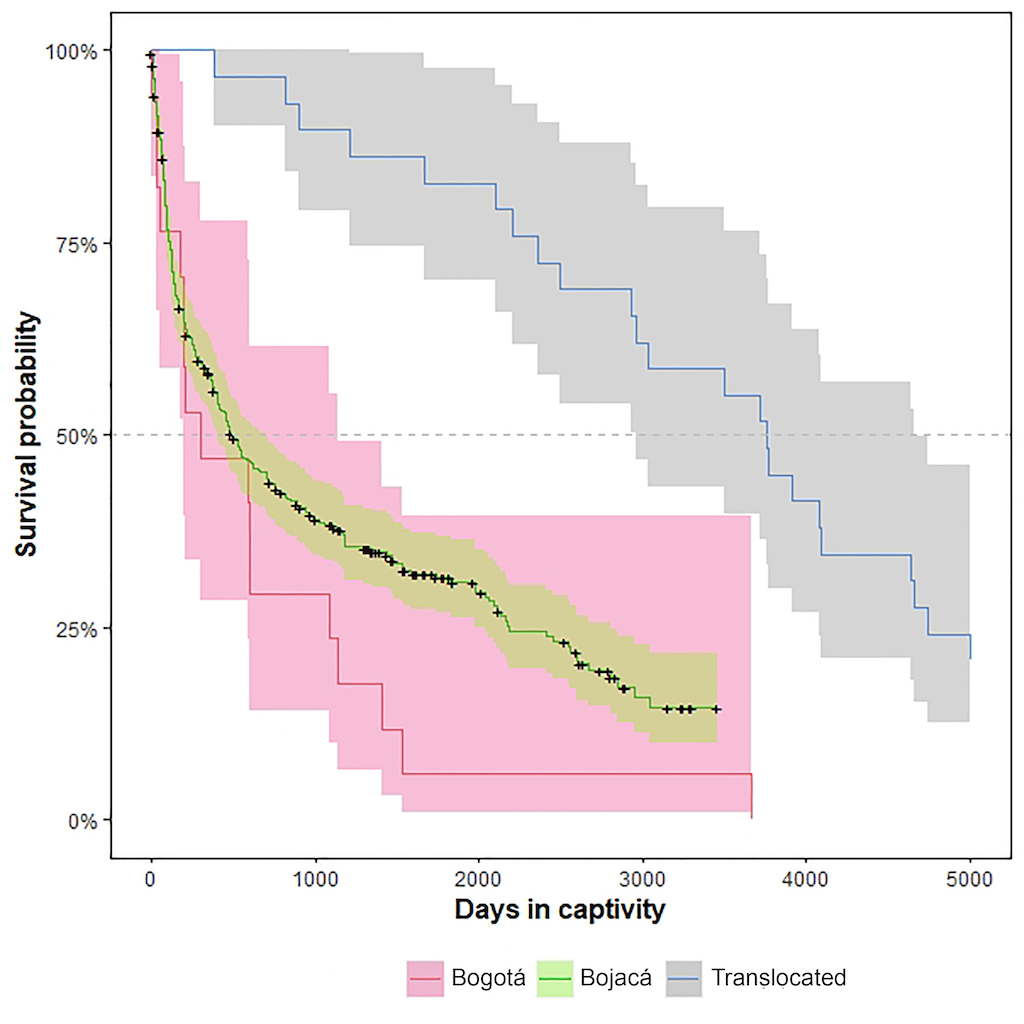
Figure 4. Median survival time of the snakes housed in the INS serpentarium facilities of Bogota, Bojaca, and snakes translocated between facilities.
Survival rates were significantly different between INS serpentarium facilities as well as in translocated snakes (X 2= 31.1, df= 2, P <0.0001). The historical decision to relocate the serpentarium from Bogota to Bojaca clearly shows that it was a success, representing early victory for establishing suitable large-scale antivenom production. Indeed, the median lifespan of the captive snake population was increased in 260 days, allowing at least four to six milking of venoms per specimen. Interestingly, snakes translocated from Bogota to Bojaca exhibited a great increase in lifespan with a median survival time of 3870 days (Figure 4). Most of the translocated specimens of Crotalus durissus (South American rattlesnake) indicated that Colombian populations of this species, had a low and steady decay exhibiting high survival rate once the adaptation period to captivity was exceeded (see below).
Over the last decade, survival rates of the captive snake populations from the INS serpentarium exhibited different rates according to their origin, family, genus, and species. In general, the median survival time that represents the time in which 50% of the captivity snake population has died, was 580 days (Figure 5A). This means that during the last decade, every 1.6 years a population turnover occurred. Differences in the median survival time in the captive snakes according to their ecoregional origins were also observed (X 2= 30.9, df= 5, P <0.0001). These significant differences were expected due the complex Colombian geography and topography that clearly configures ecoregions with their specific biotas and climates [16]. Moreover, climatological and biophysical changes have impacts on the overall performance of wild and captive snake populations, particularly for the survival rates [13,17].
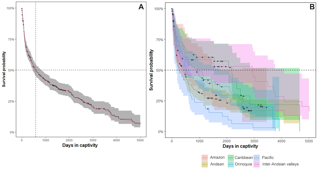
Figure 5. Median survival time of the snakes housed in the serpentarium of the Instituto Nacional de Salud during the period 2010-2021. (A) Median survival time of the entire snake population (B) Median survival time of the entire snake population by ecoregion.
Figure 5. Median survival time of the snakes housed in the serpentarium of the Instituto Nacional de Salud during the period 2010-2021. (A) Median survival time of the entire snake population (B) Median survival time of the entire snake population by ecoregion.
Species from highly humid environments like the Pacific, Amazon, and Andean regions are prone to show maladaptation syndrome that results in smaller median survival times than snake species from dry or moderately humid environments like the Caribbean, Orinoquia, and Inter-Andean valleys (Figure 5B). Maladaptation syndrome or physiological stress occurs mainly because the environmental conditions of the serpentarium are homogenous for all species despite their region of origin (temperature: 26±0.2 °C; humidity relative (Hr): 65±5), with minor changes across the serpentarium room (vertical variation ~ 3.52°C/ 5.65 Hr; horizontal variation ~ 1.52°C/ 2.65 Hr).
Therefore, this can cause significant stress on the snakes from highly humid environments, reducing the survival probability. Particularly, some pathologies observed are associated with maladaptation syndrome or physiological stress on the snakes from highly humid environments such as dysecdysis (abnormal pattern of skin shedding), mycosis (fungal infection), dermatitis (skin irritation), and dehydration [18,19]. About 50% of the snakes housed in the INS serpentarium, irrespective of their regional origin, have a median survival time of over 1 year and an average of 2.96 years for the entire snake population, indicating that the snakes in captivity for antivenom production during the last decade have been maintained under good captive conditions (Table 2).
Table 2. Median survival time and lifespan observed during the period 2010-2021 in captive snakes for antivenom production housed at INS serpentarium.
N: Sample size. LCI. Lower confidence interval. UCI: Upper confidence interval. *: Data excluded due the small sample size.
Differences of the median survival time between elapid and viperid snakes are notable (X 2= 36.6, df= 1, P <0.0001), showing that elapid species exhibit lower median survival times than viperid species (Table 2). Thus, coral snakes are more prone to show maladaptation syndromes than do viperids (Figure 6A). Comparatively, elapid species require more specialized care and environmental conditions than viperids for factors such as types of substrates, natural prey, feeding techniques, milking schedules, and handling techniques [9–11,20–24]. In addition, the lack of environmental complexity or enriched environments have been proposed as sources of stressors that directly impact the survival rates of snakes in captivity [9].
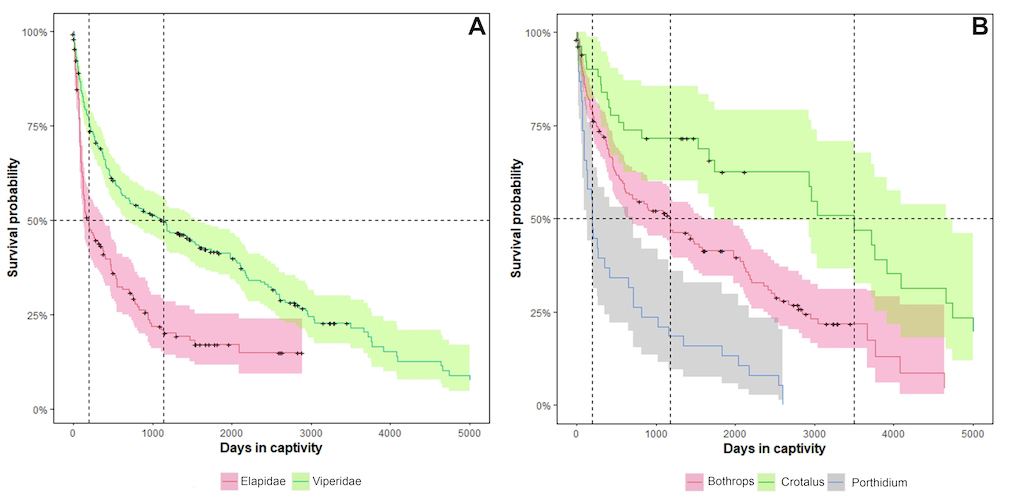
Figure 6. Median survival times. (A) Comparison between the two main families of medically important snakes housed in the serpentarium of the Instituto Nacional de Salud during the period 2010-2021 (B) Comparisons between viperid genera housed in the serpentarium of the Instituto Nacional de Salud during the period 2010-2021.
Nevertheless, median survival time observed in Micrurus species housed in the INS serpentarium overcome those reported for other serpentarium for antivenom production [23,25], indicating that the environmental enrichment done provides proper welfare for these snakes (see below). Additionally, no significant differences in the median survival time were observed between Micrurus species (X 2= 3.0, df= 1, P =0.08). Lifespan profiles were very similar between Micrurus dumerilii and M. mipartitus,in which median survival time differed by 91 days (Figure 7A).
In contrast, Colombian pitviper species seem to have a higher adaptive capacity to captive environments, but they do show significant differences between genera (X 2= 47.8, df= 2, P <0.0001). Crotalus durissus exhibit the longest lifespan and median survival time, followed by Bothrops and Porthidium species (Figure 6B). Likewise, significant differences between pitviper species are also observed (X 2= 54.2, df= 3, P <0.0001), exhibiting a distinctive lifespan profile, as well as a median survival time by species (Figure 6B). Crotalus durissus shows the highest median survival time (3,499 days), followed by Bothrops atrox (2,103 days),B. asper (891 days), and Porthidium lansbergii (198 days).
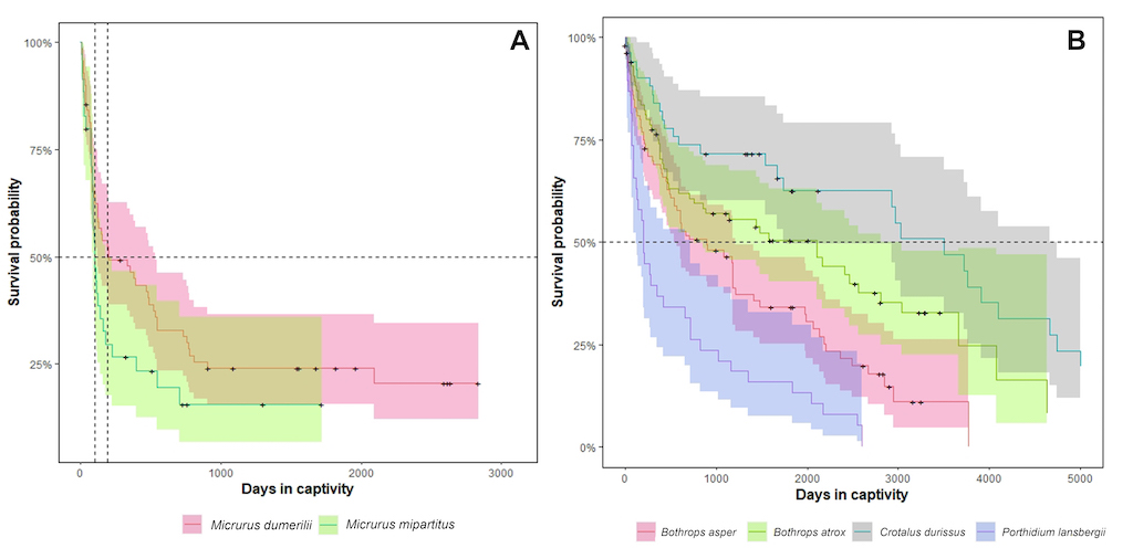
Figure 7. Median survival time. (A) Comparison between the two main medically important elapid species housed in the serpentarium of the Instituto Nacional de Salud during the period 2010-2021 (B) Comparisons between the main medically important viperids species housed in the serpentarium of the Instituto Nacional de Salud during the period 2010-2021.
In fact, Porthidium and Lachesis species (bushmasters) show similar lifespans in captivity as observed in Micrurus species, indicating that these pitvipers also require specialized care and environmental conditions. They are more likely to show the most extreme maladaptation signs or physiological stress among all viperids species housed in INS serpentarium (Table 2). Interestingly, both genera represent body size extremes seen across all the viperid species that inhabit South America; the Porthidium species was the shortest and the two Lachesis species were the longest and the heaviest, suggesting that body size plays a key role in the ability of snakes to survive in captive environments.
There are few previously documented data assessing snake survival of venomous species maintained in captivity for antivenom production in Central and South American. Nevertheless, the lifespan profile and median survival time by observed species has close similarities to previous studies done in Costa Rica, although there are clear differences. For example, Sasa et al. [23] in the serpentarium of the Instituto Clodomiro Picado (ICP) from Costa Rica, stated four statistically distinct survival profiles that closely resemble the survival curves depicted by the captive snake population of the INS serpentarium. Thus, following the Sasa et al. [23] proposal, the statistically distinct survival profiles observed in the INS serpentarium match the following categories (Figure 7B):
-
High survival —. Snake species with over 70% survival during the first 24 months (720 days) and less than 40% of deaths occurring after the 60th (1800 days) month in captivity. This profile was exhibited only by
Crotalus durissus.
-
Steady decay —. Snake species in which less than 50% of the deaths occur during the first 24 months in captivity, and a gradual reduction of survival follows. This profile was shown only by
Bothrops asper andB. atrox.
-
Rapid decline —. Snake species in which close to 40% survive during the first 24 months (720 days) in captivity but mortality rate decreases after 48 months (1440 days). No species followed this profile.
-
Severe decline—. Snake species in which close to 30% survival occurs within the first 24 months (720 days) and a steady decrease observed afterward. This profile was exhibited by
Micrurus dumerilii, M. mipartitus,and Lachesis acrochorda, and
Porthidium lansbergii.
Likewise, in the ICP snake population reported by Sasa et al [23] the median survival time of Porthidium nasutum and Lachesis stenophrys (the closest relative of Porthidium lansbergii,Lachesis acrochorda,and L. muta, respectively) showed a rapid decline in the survival curves, as well as the shortest median survival times, similar to that observed in captive populations of their Colombian congeners. In addition, both Costa Rican species also represent the body size extremes [23,26].
In the case of Bothrops asper,the single species shared by both serpentarium, the median survival time seen had marked differences, at 2.3 times longer in the Colombian rather than the Costa Rican captive snake population. In addition, both Bothrops asper captive populations showed distinct survival patterns, in which the Colombian population exhibited a steady decay of its survival profile while Costa Rican population showed a rapid decline in the survival profile.
Differences observed between ICP and INS captive snake populations of Bothrops asper can be caused by intrinsic and extrinsic factors related to captive conditions or natural history traits of each species, as well as population variability. For example, previous survival studies of coral snakes in captivity have found significant associations to the initial body condition during admission in the serpentarium, as well as to the types of substrates, and feeding schemes during captivity [21,22]. In Colombia, captive populations of the coral snake Micrurus mipartitus (redtail coral snake) have shown that weight and body condition at admission play a significant role in the lifespan observed [27].
Table 3. Cox proportional hazards models. The table depicts the P-values of each model, indicating the statistical significance of each covariate. Snout ventral length (SVL), weight, synergetic pathologies, ectoparasites, dysecdysis, and dehydration were included as intrinsic snake covariates during admission in the INS serpentarium. Synergetic pathologies illustrate the cases when a snake exhibited two or more pathologies at the same time (e.g., ectoparasites + dysecdysis)
|
Analysis categories
|
Groups
|
|
N
|
SVL
|
Weight
|
Synergetic pathologies
|
Ectoparasites
|
Dysecdysis
|
Dehydration
|
|
Regions
|
Amazon
|
|
28
|
0.387
|
0.0351
|
0.192
|
0.224
|
0.044
|
0.022
|
|
Andean
|
|
143
|
<0.001
|
0.0926
|
0.0049
|
0.445
|
0.425
|
0.135
|
|
Caribbean
|
|
64
|
0.52
|
0.99
|
0.273
|
0.531
|
0.187
|
<0.0001
|
|
Orinoquia
|
|
67
|
0.033
|
0.0111
|
0.968
|
0.796
|
NA
|
NA
|
|
Pacific
|
|
4
|
0.047
|
0.025
|
0.277
|
0.107
|
NA
|
0.909
|
|
Inter-Andean valleys
|
|
31
|
0.13
|
0.097
|
NA
|
NA
|
NA
|
NA
|
|
Families
|
Elapidae
|
|
101
|
0.0068
|
0.736
|
<0.0001
|
0.423
|
0.0167
|
0.916
|
|
Viperidae
|
|
209
|
0.245
|
0.265
|
0.429
|
0.203
|
0.06
|
0.0028
|
|
Viperid species
|
Bothrops asper
|
|
70
|
0.701
|
0.867
|
0.703
|
<0.0001
|
0.131
|
0.0085
|
|
Bothrops atrox
|
|
36
|
0.0147
|
0.0054
|
<0.0001
|
<0.0001
|
0.0001
|
1
|
|
Crotalus durissus
|
|
20
|
0.884
|
0.976
|
0.15
|
NA
|
0.99
|
1
|
|
Micrurus species
|
M. dumerilii
|
|
66
|
0.0092
|
0.931
|
0.0011
|
0.381
|
0.019
|
0.861
|
|
M. mipartitus
|
|
31
|
0.265
|
0.69
|
0.171
|
NA
|
0.63
|
NA
|
|
P-value color scale
|

|
NA: No data available. P-value color scale illustrating the statistical significance of the local differences between Cox proportional hazards models and intrinsic snake covariates.
We focused on the intrinsic factors of the conditions of snakes at admission in the INS serpentarium, such as body size, weight, and the presence/absence of pathologies, ectoparasites, dysecdysis, and dehydration. Cox proportional hazard models (CPH) indicate that the main pathologies observed during the last decade in the INS serpentarium can act alone or synergistically. In general, survival of captive snakes showed significant correlation with the intrinsic covariates assessed (Table 3). Body size (SVL) was the most frequent explanatory variable and was significant in 46% of the snake groups assessed, followed by dysecdysis (38%), weight, synergetic pathologies, dehydration (31% each one), and ectoparasites (15%). These results were expected since body size and weight loss are common risk factors in captive animals [28].
Dehydration showed a marked correlation according to the ecoregional origin of the snake or its taxonomic family. It indicates that when this sign appears, it alone has a significant effect on the probability of survival of viperid snakes from Amazonian and Caribbean ecoregions. In contrast, when dehydration appears together with synergetic pathologies, the cluster of factors shows a significant correlation of snake species from the Andean region, particularly for Micrurus dumerilii.
This finding is interesting because, as above, this same snake population is prone to show a maladaptation syndrome exhibiting smaller median survival times, suggesting that synergistic interactions could modulate or reduce snake lifespans in the serpentarium. Synergistic interactions of two or more pathologies are an important issue during snake maintenance in captivity because they can generate a greater deleterious response than when the pathologies act alone. Unfortunately, there is a dearth of information about this topic in literature, thus the understanding and the state of knowledge of synergistic pathological interactions in South American tropical snake in captivity is still extremely fragmentary.
Survival of captive snakes showed a significant correlation with dysecdysis, particularly with snakes of both viperid and elapid species from the Amazonian ecoregion. Unlike synergistic pathological interactions, this pathology is well- known and is considered as an important risk factor in captive reptiles because of multifactorial causes, such as: (1) inadequate captive management due to unsuitable humidity or temperature conditions; (2) scars from old injuries, surgery, or burns; (3) parasitic infections and systemic disease; and (4) underlying disease causing dehydration or impaired movement (e.g., fused vertebrae) [29].
Nevertheless, each snake group that we assessed exhibited a unique combination of explanatory variables with few commonalities among the CPH models. This suggests that future studies must be performed focusing on explored variables such as origin, locality/ecoregion, cortisol level, endoparasites, etc. For example, medically important snake species from the Orinoquia and Amazonian regions share body size and weight as explanatory variables. This would be expected because both regions encompassed the historical distribution of Lachesis muta, a species that represents an extreme body size among South American pitvipers.
3. Understanding the deaths of snakes in captivity
Since 2010, all deceased snake in captivity have been examined using a standardized protocol in the INS serpentarium to understand the causes of death. Results of necropsies have been documented and entered into each specimen’s clinical record. The compilation of these records year by year provides the basis of information allowing us to understand the pathologies and their impacts on the general population in the serpentarium. Understanding the causes of death of snake in captive conditions provides an important input in the improvement of management and tenure techniques in a large-scale snake captivity program for antivenom production [23,30].
The following descriptions and analyses were based on 264 necropsies done by veterinarians from 2010 to Jan of 2021 in deceased captive snakes encompassing 99 elapids (M. camilae, M. dissoleucus, M. dumerilii, M. filiformis, M. helleri, M. hemprichii, M. medemi, M. mipartitus, M. nattereri, M. obscurus, and M. surinamensis) and 165 viperids (Bothriechis schlegelii, Bothrocophias tulitoi, Bothrops asper, B. atrox, B. bilineatus,B. venezuelensis, Crotalus durissus, Lachesis acrochorda,L. muta, and Porthidium lansbergii; Table 4). Mostly, necropsy records came from the INS serpentarium since 2012 from the Hacienda Galindo in the municipality of Bojaca, Cundinamarca (see Chapter 7).
Digestive and respiratory system abnormalities were the main findings. They represented 43.3% of the total postmortem findings, followed by an absence of abnormalities (Figure 8). Digestive abnormalities in snakes are the main cause for infections. However, due the lack of established diagnostic tests, references for most reptile species and correct interpretation of the clinical signs are challenging [31,32]. Research efforts of diagnostic techniques, particularly imaging (e.g., contrast radiography and ultrasonography) of gastrointestinal tract, have resulted in clinical advancements for practicing veterinarians [32].
Nevertheless, due to limited technical facilities in serpentariums dedicated to antivenom production, as well as the dearth of reptile veterinarians available in Colombia that could to provide an accurate diagnosis, the therapy of digestive system disorders in captive snakes is difficult [31]. Therefore, in both pet veterinary clinics and laboratory animal production units like the INS serpentarium, digestive diseases are common as a cause of death; but this cause mostly cannot be detected until postmortem examination. Currently, there are no feasible factors or enclosure conditions that can be linked to a snake’s death nor any digestive system abnormalities that have been observed in the INS serpentarium. This finding requires further research.
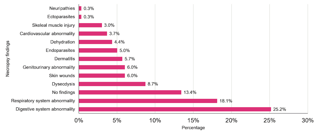
Figure 8. Main postmortem findings in medically important snake species housed in the INS serpentarium during 2010-2021.
Respiratory diseases in snakes have multifactorial origins, including bacterial, viral parasitic, fungal, stress, cage spacing, genetics, physiologic ecdysis, etc.[33]. Respiratory diseases are common in captive snakes since enclosure design and space directly impact the health of the snake. These factors affect the normal behavior and mobility of snakes, as well as the additional captivity conditions like sanitation and humidity or physiological conditions like thermoregulation [28]. An ideal enclosure should provide a dry, hot area; a warm, humid area; a cool, dry area; and a cool, damp area [28]. However, as mentioned earlier, due to the limited space available in the INS serpentarium, all enclosures share the same environmental conditions and are homogeneous, lacking a diverse thermal offering. Therefore, prolonged exposition to invariant thermal and humidity conditions in the captive environment could explain the abnormalities observed. However, continued research efforts must be done to assess this hypothesis.
Table 4. Main causes of death in medically important snake species housed in the INS serpentarium during 2010-2021.
N= sample size. BBCA= Bad body condition before admission.
Knowledge of the diseases that snakes contract and the microbial agents they carry is essential for their management in captivity [34,35]. But there are few scientific studies that focus on the diseases of South American tropical snakes used for antivenom production. Nevertheless, the necropsy reports from the last decade of the INS serpentarium showed close similarities to previous observations in captive snakes or zoological exhibitions of specimens [13,18,29,30].
For example, the five most common causes of death in captive snakes in order of importance are (Table 4) as follow:
-
Unknown causes.— Defined as the impossibility of establishing a medical cause of death from necropsy findings. This cause groups the largest proportion of snake deaths in all previous studies [13,18,29], representing 53% of deaths in the INS serpentarium with a notable prevalence in viperid species.
-
Maladaptation syndrome.— Prevalence of 32.2% observed in captive snakes in the INS serpentarium, exhibiting an apparent association with body size and ecoregional origin, with significantly high frequency in Micrurus species.
-
Poor body condition.— Deaths resulting from a deficient body condition before admission. The observed frequency of cases in the INS serpentarium was 10.61%. This cause is highly relevant during the first 150 days of captivity for all species but presents a high frequency in Micrurus species.
-
Accidental death.— Deaths resulting from an accidental event in captivity. This cause had a low prevalence (2.27%), with mechanical asphyxia produced by attempts to escape from the enclosure being the main accident.
-
Iatrogeny.— Deaths related to illness or injury caused during medical examination or treatment. This cause had the lowest frequency (1.89%) among observed causes of death in the INS serpentarium. However, when it occurred, coral snakes were the most prone to death from medical procedures.
4. Learned lessons
4.1 Lesson one: Feeding and nutrition
One of the main concerns for maintaining captive snakes for antivenom production is providing suitable feeding and nutrition; the goal is to prolong a healthy life for the animal to the extent possible. Feeding constitutes one of the most important and decisive activities for the maintenance and survival of snakes in captivity. Most captive snakes prefer live prey or prey that has been freshly killed, although they also accept dead prey [36]. The latter option has been commonly adopted by snake keepers in zoos or educational biological exhibitions, but dead prey is not compatible with good manufacturing practices in a program for antivenom production.
To achieve international production standards, special procedures must be designed, considering the following aspects: (1) A suitable selection of the feeding technique according to the diet type for each snake species should be chosen; (2) The establishment of a feeding schedule according to the needs of the snake including the following criteria: health status, body size, body mass index, developmental stage (neonate, juvenile, adult); and (3) Feeding procedures performed by an experienced team of snake keepers.
Most feeding and nutritional problems arise from improper care or handling, combined with poor feeding management [37]. This lesson summarizes the main conclusions gained from solving feeding and nutritional problems that arise in the INS serpentarium housing tropical snakes that seek to minimize deaths and diseases caused by malnutrition.
Voluntary feeding
As a primary directive, snake keepers must maintain and promote as long as possible the voluntary feeding stimuli exhibited by the snakes when they entered the serpentarium from wild environments. Steady voluntary feeding stimuli avoids the occurrence of a maladaptation syndrome. For success, feeding procedures must be performed during the day in a warm enclosure with temperatures ranging between 26° to 30°C and providing live prey. Both elapids and viperids tend to feed using this approach; however, technical steps differ. The technical approach employed to feed viperids snakes is as follows (Figure 9):
- The snake keeper must remove the snake from its enclosure and put it in a box for weighing; this allows a determination of the prey to be given. This procedure should be done previously to the following steps.
- The snake keeper selects a rodent according to the snake’s weight (~ 10% of the snake’s weight).
- The rodent is held by a long tweezer and offered to the snake using the lid of the enclosure as an additional security barrier. Displaying gentle movements at 10 to 20 cm from the head of the snake, the snake keeper seeks to stimulate the snake to bite or attack the prey. The distance at which prey is offered to snakes varies according to the body size of the snake; we recommend displaying gentle movements when offering the prey at distance not more than 30% of the snake's body length.
- Once the snake bites or attacks the prey, the prey is released from the tweezer and the enclosure is closed. If the snake refuses to bite the prey it is withdrawn, and an offering of prey is put off until the second round of feeding. The prey must be removed to avoid psychological stress or wounds caused by the prey. Attacks by the prey can induce a severe, potentially life-threatening infection. The prey is never released without confirmation of an effective bite by the snake and the prey is dead.
- In the case that more than one prey is needed to reach 10% of the required feeding weight, the snake keeper must wait for a second round of feeding, checking that the snake has completely swallowed the first rodent; then the additional prey is offered. In the event of a delay in the consumption of the first prey, the snake keeper performs euthanasia on the second prey, and it is left inside the enclosure. Ingestion of the prey by the snake must be confirmed, otherwise it must be removed.
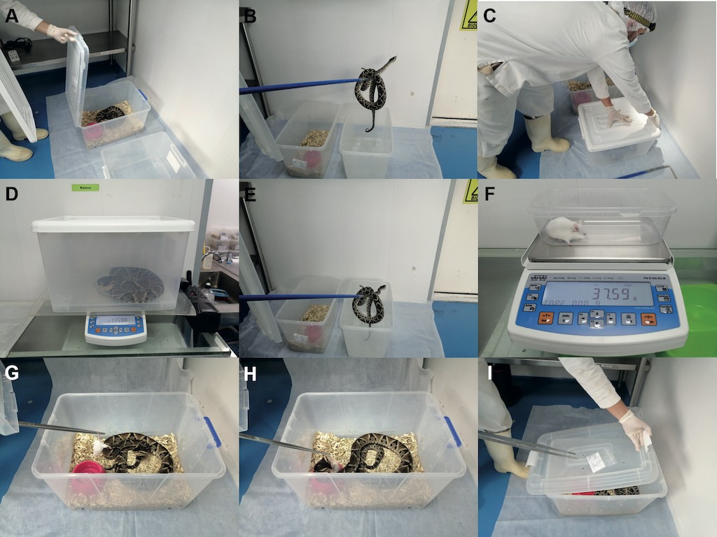
Figure 9. Weighing and feeding viperids. (A) Opening the snake enclosure using the lid as protective barrier. (B) Using a snake-hook, shifting the snake to an empty box for weighing. (C) Closing the box in which the snake will be weighed using the same lid of the snake enclosure. (D) Weighing the snake on a scale. (E) Returning the snake to its enclosure and closing the box. (F) Weighing the rodent on a scale. (G) The mouse is offered to the snake, holding the prey by a long tweezer, and displaying gentle movements. (H) The snake bites or attack on the prey; the prey is released from the tweezer. (I) The enclosure is closed.
The INS has an animal house facility independent of the serpentarium, used to produce certified rodents raised in controlled conditions, providing a suitable diet free of pathogens for viperid snakes in captivity (Table 5). Thus, the INS serpentarium always has broad dietary offerings varying in size and weight as food for the snakes according to their needs.
Table 5. Rodents raised in controlled conditions used to feed viperid snakes in the INS serpentarium.
|
Type
|
Strain
|
Strain code
|
Fur color
|
|
Mice
|
BALB/c
|
028
|
Albine
|
|
Mice
|
CD1 (ICR)
|
022
|
Albine
|
|
Rat
|
Wistar
|
003
|
Albine
|
|
Guinea pig
|
Hartley
|
051
|
Albine
|
|
Gerbil
|
Mongolian
|
243
|
Dark
|
|
Hamster
|
Syrian
|
249
|
Golden
|
Unlike viperids, feeding coral snakes with live animals has enormous constraints and technical issues that reduce the possibility of steady voluntary feeding stimuli. First, due to the fact that coral snakes are ground and litter dwellers (semi-fossorials), their diet consists mainly of other snakes, limbless lizards, caecilians, swamp eels, and knife fish [38]. In practice, the collection, maintenance or raising in captivity of this kind of prey is very difficult and unsustainable. So, feeding coralsnake with live animals in the context of international production standards for snake antivenom production is not feasible or it is highly expensive. Second, due to such prey being collected from the wild, it is not possible to guarantee they are free of pathogens; and the prey would be very difficult to sanitize. Third, providing a continuous feeding schedule according to snake needs based on prey collected from the wild is difficult to achieve.
So, in practice feeding coral snakes with live animals is an occasional and opportunistic activity. The object is the promotion of voluntary feeding and avoiding direct handling that has deleterious effects on the snakes’ health if force feeding is performed. At the INS serpentarium voluntary feeding with live animals is occasional. Voluntary feeding has been performed successfully in some individuals of Micrurus dumerilii, M. mipartitus, M. helleri, M. sangilensis and M. hemprichi, fed with live prey to snakes of the genera Ninia, Atractus, Oxyrhopus and Stenorrhina and limbless lizards of the genus Bachia. The technical approach employed to feed elapids is as follows:
- The snake keeper must remove the snake from its enclosure and put it in a box for weighing that allows the determination of the prey to be administered. This procedure should be done previously to the following steps.
- The snake keeper selects a prey according to the snake’s length and weight (less than 50% of the snake’s length and ~ 10% of the snake’s weight).
- The prey is released live into the enclosure and afterwards it is closed. The snake keeper must make the rounds verifying that the snake has attacked and ingested the prey. If it does not ingest the prey, the prey must be removed. In this way, the coral snake is stimulated to forage by using sight, smell, and taste.
- The consumption of the prey must be confirmed within a period of two days, otherwise the prey must be removed.
Force feeding
Force feeding refers to the provision of mechanically forced food. This procedure is practiced by snake keepers when snakes refuse prey repeatedly for more than four months, due to the prevalence of some medical condition or when operative constraints when feeding with live animals is not feasible. This procedure can be performed in two ways. First, using a tweezer, the entire prey is introducing manually into the digestive tract of the snake, avoiding any kind of restrictive tool to hold the snake (e.g., restraining tube). The prey must be selected according to snake’s length and weight as described above. Second, by introducing an orogastric probe into the digestive tract of the snake and gently injecting a nutritive formula. Following, both techniques are described.
Force feeding using a whole prey.— According to the accumulated experience of the INS serpentarium, this technique is mostly used in viperids rather than in elapids because the latter are prone to be highly stressed by handling during feeding procedure. Most viperids endure handling without major difficulties during force feeding. The technical approach employed to feed snakes using a whole prey is as follows:
- The snake keeper must remove the snake from its enclosure and put it in a cloth bag for weighing. This allows determination of the prey to be administered. The procedure must be done between five to ten days before feeding.
- The snake keeper selects a prey according to the snake’s weight (~ 10% of the snake’s weight).
- The euthanized prey is moistened with water for lubrication.
- When catching and gently handling the snake to be fed, it is recommended that the following steps be conducted by two snake keepers. The first snake keeper is charged with capturing and holding the snake, the second keeper is in charge of performing the feeding. Avoid the use of any kind of restrictive tool to hold the snake during feeding procedure (e.g., restraining tube).
- A long tweezer (at least 20 cm) is used to hold the pre-lubricated prey with the tips of the tweezer.
- Insert the pre-lubricated prey (headfirst) into the mouth of the snake.
- With gentle and slow pushes, the pre-lubricated prey is pushed into the digestive tract of the snake, passing the head till a distance equivalent to the snake’s head-length is reached. Afterwards, release the prey from the tweezer.
- With the free hand, the snake keeper executing the feeding technique, gently massaged the ventral surface of the snake, slowly displacing the prey till reaching approximately the end of the first third of the snake’s body.
- Releasing the snake into its enclosure. Verify the acceptance of the prey for 24 hours. In case of regurgitating the prey, the prey must be discarded.
Force-feeding using a nutritive formula.— This technique represents one of the greatest successes of the INS serpentarium in the last decade. The use of a nutritive formula as alternative force feeding allowed us to consolidate a steady Micrurus snake population exceeding the average lifespan 1.4 years and a median survival time of 91 days, including captive specimens with a lifespan of 8 years.
The accumulative experience teaches us that the two main success factors were: (1) The composition of the nutritive formula; and (2) the technical improvement over time that encompassed the experience gained by the snake keepers in handling snakes, introducing a Nelaton catheterinto the digestive tract, and accurate performance and following feeding steps using the nutritive formula.
The development of the nutritive formula was the response to the strong constraints to obtaining prey from the wild that allow a continuous feeding schedule according to coralsnake needs, as well as the frequent rejection of live prey in the first months of captivity. The nutritive formula is a hyperproteic solution based on “criolla” chicken eggs (criolla = a hen raised in a natural way in the countryside) mixed with Casilan® (Composition: protein = 95 %, carbohydrates = 1%; fat = 4) plus vitamin and amino acid supplements. Based on the experience of the INS serpentarium veterinarians, the use of “criolla” chicken eggs is mandatory. The use of other kinds of eggs provoked the rejection of the nutritive formula by the snakes. Because Casilan® disappeared from the market in 2021, the nutritive formula changed, replacing Casilan® by Reptomin® (Composition: protein = 44 %, carbohydrates = 3%; fat = 5%, humidity = 9%, Calcium = 0.3%, Phosphorus = 0.4%) and observing the same previous good results using Casilan®.
The nutritive formula has shown excellent results for most of the coral snakes although this was not an infallible solution. After feeding ten Micrurus surinamensis (aquatic coral snake) with nutritive formula that replaced the “criolla” chicken eggs by fish as a source of protein, all snakes exhibited severe life-threatening effects such as dull skin, incomplete ecdysis, and immobility. Therefore, the use of a nutritive formula based on a fish-based diet was contraindicated. In the case of M. surinamensis, the nutritive formula based on water plus Reptomin® showed acceptable results, although it is still a challenge.
The preparation and administration of the nutritive formula must be careful and very liquid. Dense consistencies can make digestion difficult. Lumps or bubbles can cause the rupture of the digestive tract during administration. Handling time must be minimized by quick administration of the nutritive formula. Therefore, the introduction of the Nelaton catheter must be quick, gently pushing into the digestive tract. The gauge of the Nelaton catheter must be selected according to the snake’s body length. Handling must be done using bare hands to avoid the use of tweezers or any kind of restrictive tool that provokes stress in the snake and consequent rejection of the nutritive formula.
Consumption of the nutritive formula must be registered in the clinical record of each snake. According to accumulated experiences feeding coral snakes, the suitable proportion of nutritive formula to feed snakes is between 10% and 30% of the snake’s weight, considering a proportion 1:1 (body weight g/formula mL). However, the proportion of the nutritive formula supplied can vary between individuals because the stage development, medical condition, or body condition of the snake. Clinical veterinary records must be consulted, looking to adjust the percentage according to the historical records of each specimen.
Three snake keepers must perform force-feeding when using the nutritive formula. The technical approach employed to feed snakes using nutritive formula is as follows (Figure 10):
- The snake keepers must remove the snake from its enclosure using a narrow herpetological hook and put it in a cloth bag for weighing to allow an estimate of the volume of formula to be administered.
- Preparation of a Nelaton catheter and syringe according to snake body size with nutritive formula (A 3mL syringe with Nelaton catheter of 6 gauge for snake body size less than 20cm; a 20mL syringe with gavage tube of 9 or 10 gauge for snake body size more than 20cm).
- Performing force-feeding using nutritive formula handling must be done using bare hands, avoiding the use of tweezers or any kind of restrictive tool. The first snake keeper is in charge of capturing and holding the snake, the second keeper takes care of the feeding technique.
- The first snake keeper introduces the Nelaton catheter up to the first third of the snake’s body. It must be introduced with a gentle but quick push into the digestive tract.
- Immediately after the introduction of the Nelaton catheter, the second snake keeper must introduce the controlled supply of the formula, while a third snake keeper holds the snake’s body, performing gentle massaging on the ventral surface of the animal. A steady injection speed of the formula through the syringe must be maintained, avoiding a buildup and bubbles that could provoke indigestion or digestive tract obstruction.
- Once the entire volume of nutritive formula has been administered, the Nelaton catheter is slowly withdrawn from the digestive tract. The snake’s mouth must be washed with water before the snake is returned to its enclosure.
- Returning the snake to its enclosure. Verify the acceptance of the nutritive formula for 24 hours. In case of regurgitating the formula, clean the enclosure and reprogram a feeding procedure.
Note: This feeding procedure cannot be carried out in specimens that are experiencing ecdysis unless the veterinarian suggests it is ok to do so. If any additional medical procedure is necessary, it must be carried out before the feeding procedure.
The established feeding schedule in the INS serpentarium is monthly for viperids and fortnightly for elapids. Nevertheless, neonates of both viperid and elapid snakes feed each 10 and 15 days due to their high metabolic demands. Other reasons for the change in feeding frequency are due to specific veterinary medical decisions. The feeding schedules, having been established over 10 years, exhibit good performance and results. However, we recommend that each serpentarium according to the species of snake housed must be assessed for the correct feeding frequency.
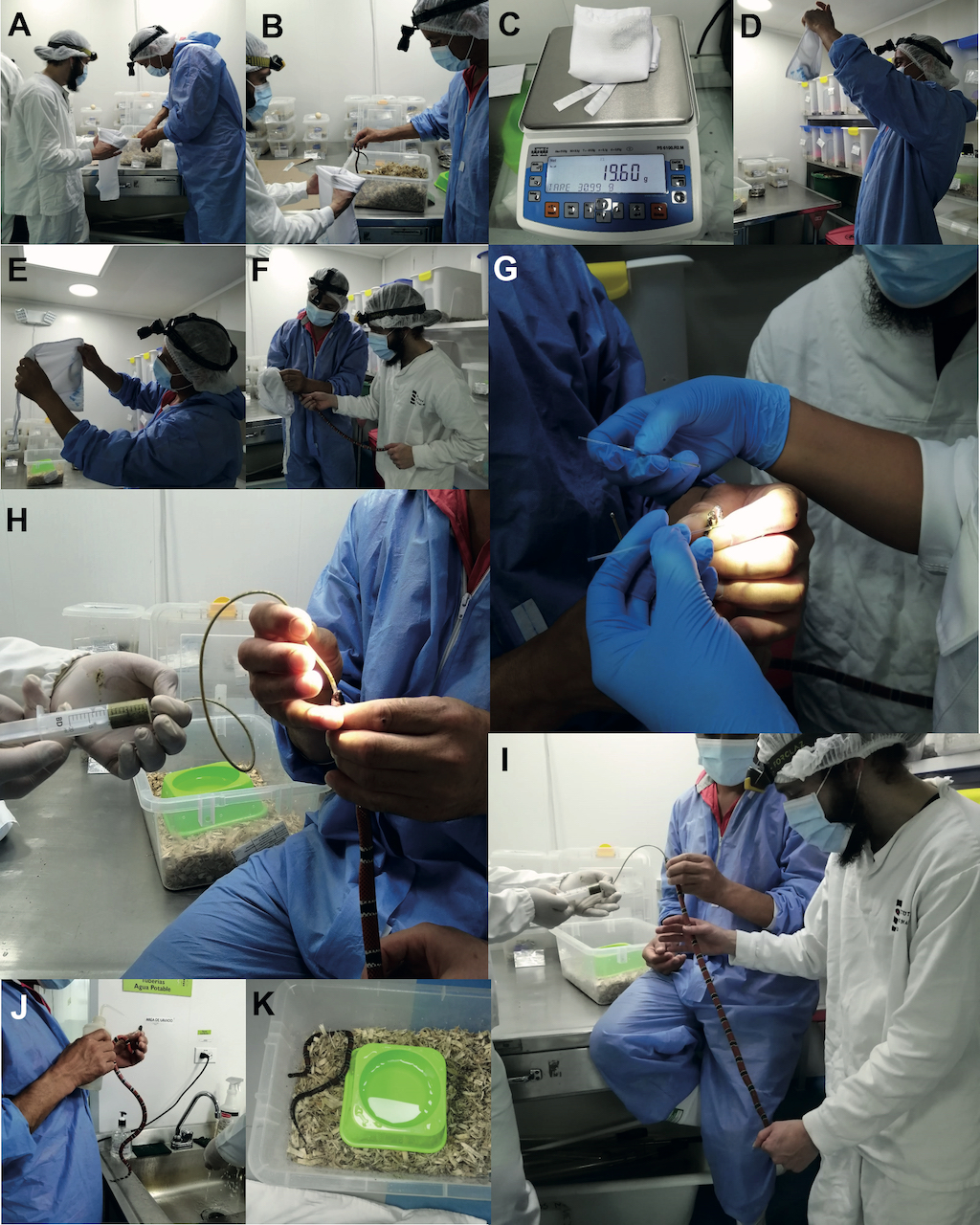
Figure 10. Weighing and feeding process of Micrurus species. (A) Opening the snake enclosure using the lid as protective barrier. (B) Using a narrow herpetological hook to shift the snake into an empty white cloth bag. (C) Folding the white cloth bag and weighing the snake. (D-E) The first snake keeper, observing the snake against the light, locates the snake head. (F) The first snake keeper immobilizes the snake´s head while the second snake keeper gently holds the snake’s body. (G) A third snake keeper milks the venom using two capillary tubes. (H) The first snake keeper introduces the Nelaton catheter up to the first third of the snake’s body. Insertion must be with a gentle but quick push into the digestive tract. (I) The second snake keeper begins introducing the controlled supply of the formula, while a third snake keeper holds the snake body while performing a gentle massage on the ventral surface of the animal. (J) The Nelaton catheter is slowly withdrawn from the digestive tract. The snake’s mouth must be washed with water before being returned to its enclosure. (K)The snake is then returned to its enclosure.
4.2 Lesson two: Iatrogenic deaths
Despite iatrogenic deaths representing less than 2% of snake deaths in the last decade in the INS serpentarium, it is important to describe the most relevant events as learned lesson to be considered in future implementations of a large-scale program for antivenom production based on captive snakes.
Case 1: digestive tract rupture.— Force-feeding using nutritive formula through a Nelaton catheter is an adapted technique from those feeding techniques used to feed murine models in research procedures [39]. Thus, there are no references of its use in snakes, and therefore our learning was done through experience. Force-feeding employing a Nelaton catheter requires an experienced snake keeper for employment; meanwhile experience is gained, mistakes arise, and the most common mistake results in digestive tract rupture.
The main factors that contribute to digestive tract rupture are the following: (1) unsuitable selection of the Nelaton catheter size that mismatches the snake body size; (2) an inadequate entrance of the Nelaton catheter that exceeds the first third of snake’s body; (3) an overly rapid and too strong push entering the Nelaton catheter; (4) an uncontrolled supply of the nutritive formula that generates a buildup and bubbles; (5) lack of a gentle massage on the ventral surface of the snake; and (6) fast and uncontrolled extubating. Based on our experience, death by digestive tract rupture can be detected within the first 72 h after the force-feeding procedure.
4.3 Lesson three: environmental enrichment
Environmental enrichment is the set of techniques and management measures used by keepers looking to encourage the expression of natural behaviors of animals in captive conditions [40]. Additionally, these management measures seek to promote and increment the survival, health, and well-being of captive animals [41].
Historically, environmental enrichment of snake captives has been mostly studied in zoos and educational biodioramas rather than serpentariums that are focused on antivenom production [25]. For example, for public environmental educational initiatives, Cardoso et al. [42] demonstrated that enriched environments have significantly positive effects on the welfare and behavior of captive populations of Micrurus corallinus, reducing stress and preventing maladaptation syndromes.
Environmental enrichment of serpentariums focused in intensive antivenom production is challenging due to the lack of research that addresses which are the “best” practices. Particularly, environmental enrichment needs to overcome special challenges for standardized animal sanitation protocols in the context of an intensive snake captivity program that seeks replicability and reproducibility [25]. For example, to maintain high standards of sanitation all elements (tree branches, rocks, pieces of tree trunks, plants, etc.), both natural and artificial, must be sanitized to turn them into aseptic elements for the control of pathogenetic sources. Many disinfectants are inactivated in the presence of organic matter, so washing and disinfecting trunks and plants is useless for the snake health.
An intensive snake captivity program for antivenom production that meets the requirements of good environmental enrichment is a challenging task because a high number of snakes must be cared for daily that includes the amount of time that must be invested in milking, as well as the trained staff and resources required to do so. Commonly in this kind of serpentarium there is a trade-off between environmental enrichment and operational snake management. The extremes of this trade-off are, on the one hand, an enriched biodiorama or terrarium ornamented as a faithful replica of the snake’s known microhabitat (like exhibition serpentarium), and on the other hand a basic enclosure without snake microhabitat elements, only with water supplied ad libitum.
The experience gained during INS serpentarium history suggests that the midpoint between both extremes is the “best” choice despite having different environmental enrichment needs according to species specificity. According to Loughman [43] protocol maintenance of snakes in captivity and the environmental enrichments of its enclosures should be based on the natural history of the snakes. The INS serpentarium follows this approach but adjusts it according to the INS’s own rigid and standardized operational guidelines for safety, health, welfare of animals, worker safety, product quality, and traceability. These details are similar to those stated by the Center for the Study of Venoms and Venomous Animals (CEVAP) at São Paulo State University (UNESP) Brazil [25]; and World Health Organization (WHO) guidelines for snake antivenom production [44].
Intensive serpentarium
General captive conditions.— The serpentarium houses 182 specimens, of which 146 are viperids and 36 are elapids. Each specimen has an alphanumeric code that acts as its identification number. Adults of the species that reach large body size have a chip implanted that is no larger than a grain of rice that provides an alphanumeric code. The captive environment maintains steady physical conditions (temperature: 26±0.2 C; humidity relative 65±5 humidity relative). Nevertheless, due to the area of the serpentariums space the relative humidity and temperature is not homogeneous across the room. There are zones that are more humid and other zones are drier, as well as zones that are relatively warmer and others less warm. Therefore, taking advantage of this variability snake enclosures are arranged based on the relative humidity and temperature of the original habitat of the snakes. For example, Crotalus durissus enclosures are in the drier zones of the serpentarium while Micrurus surinamensis enclosures are in the humid sections. Also, inside the enclosures the relative humidity and temperature varies, indicating that the polypropylene boxes provide a microclimate (Table 6).
Table 6. Records of thermohydrometer inside the snake enclosures
Enclosure.— Snakes are housed in individual translucid polypropylene boxes of varying sizes (20 cm long x 15 cm wide and 20 cm high used in snakes less than 30 cm SVL; up to boxes of 60 cm long x 40 cm wide and 35 cm high used for large viperids such as snakes of the genera Bothrops and Crotalus up to 2 m SVL). The advantages are safety, ease of cleaning, and maintenance. In this manner, boxes are easily monitored with relation to feeding, and health. For special cases of large snakes like Lachesis,thesize of the enclosures is 100 cm long x 45 cm wide and 35 cm high, built with stainless steel shelves with rigid translucent acrylic walls.
Substrate.— The substrate that covers the floor of the box must meet four criteria: (1) offer comfort to the snake; (2) help to maintain conditions of humidity in the enclosure; (3) facilitate cleaning of the enclosure; and (4) be deep enough to allow snakes with semifossorial habits to hide. A 1:1 mixture of shavings (poplar shavings) and an inert sanitized vermiculite (hydrous phyllosili-cate mineral) fills the floor of the box 2 cm deep, generating bedding that allows the maintenance of relative humidity at about 70%. In the case of semifossorial snakes, the floor of the box is filled up to 5 cm deep. Other substrates like natural soil, natural leaf litter, or sawdust are not recommended since they are common sources of pathogens and are difficult to sanitize. Unlike all other snake species in the serpentarium, Crotalus durissus requires the driest microhabitat, so besides placing the enclosure in the drier zone of the serpentarium, corrugated cardboard is used as a substitute of mixed vermiculite to reduce the relative humidity inside the enclosure.
Water supply.— The enclosures of adult snakes have a circular plastic drinker of 15 cm in diameter and 10 cm high, while enclosures of juvenile or neonate used have 5-ounce disposable plastic cups embedded in the substrate and attached to the floor of the enclosure. Water is administering ad libitum, and it is changed every fifteen days, however, this time may decrease depending on whether it becomes dirty due to defecating or substrate.
Enclosure ornamentation.— The arrangement of ornamentation inside the enclosures, in addition to providing an environment similar to the natural habitat, must allow for the safe handling for serpentarium staff, always reducing snakebite risk during the routine procedures of cleaning, feeding, and milking. Therefore, elements of ornamentation in the enclosures are the minimum necessary according to the natural history of the snakes. For example, the plastic water container works as a shelter for coral snakes, neonates, and juveniles of viperid species. Additional to the mixed vermiculite substrate and plastic water container, the enclosure of arboreal snakes like Bothriechis schlegelii or Bothrops bilineatus are ornamented with a set of sanitized tree branches assigned per specimen in such a way that each snake always uses the same set of branches, avoiding cross contamination.
In the case of very large snakes (over 2,000 mm snout-vent length) such as Lachesis, experience has taught us that enclosures with fallen objects help in shedding for this kind of snake. So, the enclosures are enriched with heavy wooden logs and daily water sprinkling. Both shelters and tree branches must be strategically placed to allow the snake to feel hidden while it remains visible to the serpentarium staff.
Rattlesnakes are one of the most representative species in the INS serpentarium with the highest median survival time among all species (9.7 years), as well as being one of the most numerous historically in the collection (54 specimens). No ornamented elements are used to enrich the captive environment of Crotalus durissus, only a corrugated cardboard and a plastic water container are inside their enclosure. The inclusion of elements that accumulate humidity (e.g., vermiculite, wooden logs, or plastic shelters) has been associated with pathologies like mycosis and incomplete ecdysis. Therefore, this is an example illustrating that not all snakes require complex environmental enrichment, some species need only a few elements to adapt to captivity and enjoy well-being.
4.4 Lesson four: unexpected survivors
Several snakes housed in the INS serpentarium were donations from Colombia’s Environmental Regional Autonomous Corporations (CARs, Spanish acronym), Environmental police, Fire Department, obtained through rescues, confiscation, or forfeitures. In several cases previous snake handling and captivity were improper, causing deleterious effects on the initial condition of the snake before admission to the serpentarium. Consequently, several pathologies arose significantly impacting the snake’s survival probability. Nevertheless, there are remarkable cases of injured snakes that survived for a long time, despite wounds or pathologies. This lesson illustrates the relevant events of snake survivors after improper handling procedures when the animals exhibited exceptional recovery that teaches us that, despite poor conditions of some snakes, the will to live never gives up.
Case 1: Micrurus dumerilii INSV-SR-109
Snake’s health condition during admission.— An adult female snake (SVL:680 mm; TL: 65 mm; weight: 59.8 g) from Viota, Cundinamarca, Colombia, admitted in July of 2016 presenting a musculoskeletal injury in the first third of the snake’s body, showing a skin wound with a depression or convexity possibly caused by crushing or a strike with a blunt object, causing ataxia and constraining the natural serpentine movements after the wound. The snake remained in abnormal anatomical positions in the enclosure, elongating and inverting body position (body ventral surface visible from above) after the injury, without the possibility of displaying normal behaviors such as hiding in the substrate.
Veterinary considerations in the captive management.— One month after admission, the snake presented weight loss (normal at the beginning of captivity). A structure compatible with a rib was observed protruding from the skin, exhibiting necrosis surrounding the wound. In accordance with veterinary medical criteria, the snake was kept under observation, no food was offered but water was provided ad libitum. No additional medical procedure was performed.
In the second month of captivity, a yellow coloration was observed in the rib (non-viable rib bone) protruding from the skin, showing an evident skin retention in the wound area. The skin retained was manually removed and the entire detached rib. Clotrimazole topical was applied, and the snake was returned to its enclosure without evidence of improvement in body mobility.
In the third month of captivity (October) the snake was entered into the production scheme, performing procedures of capture, handling, milking, and feeding using force feeding with nutritive formula. At this point, the snake had lost 36.8% of its initial weight (weight: 43 g).
In the following months, the snake regurgitated, rejecting the nutritive formula on several occasions, causing a dramatic weight loss until the eighth month. In the nineth month weight recovery was observed, as well as the first signs of acceptance of the nutritive formula; recovery of health and body condition reaching the admission weight approximately in May 2017 (tenth month).
The snake weight and health were recovered in the following five months, as well as acceptance of force feeding using nutritive formula, showing good adaptation to the captive environment. The wound healed without need of medical treatment or intervention, leaving a marked scare in the area of injury. However, ataxia continued without improvement in body mobility after the injury, constraining normal behavior in the enclosure (inability to hide in the substrate or artificial shelters). Due to the injury, normal ecdysis was obstructed making it necessary for the snake keepers to help in shedding.
Adaptation, survival, and death expectation.— Due the severity of the injuries the initial diagnostic was negative. Besides, according to accumulated experience with captive Micrurus species, the predicted life expectancy was less than two months, which could have decreased more after veterinary intervention to detach the rib. However, despite all expectations, the snake lived for six years (until June 22 of 2022) as part of the production scheme, including it in the routine activities of milking, and feeding.
Necropsy did not provide a conclusive cause of death. However, during the last ten months before death, the snake showed a deterioration of health, exhibiting weight loss and shedding retention in the entire body, as well as progressive general immobility, which are clear signs of aging for geriatric reptiles [45].
Case 2: Micrurus dumerilii INSV-SR-113
Snake’s health condition during admission.— An adult female snake (SVL:690 mm; TL: 82 mm; weight: 60.3 g) from San Onofre, Sucre, Colombia, admitted in September 17 of 2017, presenting good conditions of health.
Veterinary considerations in captive management.— One month after admission, the snake presented weight loss (normal at the beginning of captivity), exhibiting opacity in the internasal and parietal scales. Nevertheless, a complete ecdysis had been observed during the first month of captivity, so it seemed unlikely that the opacity was due to shedding retention. In the following months, the snake showed signs of maladaptation syndrome, such as rejection of the nutritive formula (regurgitation), excessive ecdysis (two events of shedding per month), weight loss, opacity in both head and dorsal scales.
Since the maladaptation syndrome persisted, force-feeding using nutritive formula was performed during ecdysis to attempt to stop the weight loss and depressed body condition of the snake, despite the risks generating shedding retention. Opacity in both head and dorsal scales was clinically attended as a mycotic infection; however, the treatment had no effect. Thus, scale opacity became a chronic condition of the snake. Ecdysis had to be aided by the snake keepers in each shedding event. This captive management technique was maintained for a long time and the weight loss and depressed body condition of the snake was stopped, but not the chronic signs of excessive ecdysis and scale opacity.
Adaptation, survival, and death expectation.— This case exemplifies how a snake admitted to the serpentarium to a captivity environment with good health conditions and veterinary diagnosis, quickly developed maladaptation syndrome that threatened its survival. However, thanks to suitable veterinary care and management, this trajectory was reversed, prolonging its life expectancy, despite the snake never having suitable adaptation to the captive environment. The snake lived four years in captivity and was included in the routine production scheme of the INS serpentarium till it died, though no other signs or pathologies were observed.
Appendix: Material and Methods
Based on the history of the data available from 1990 to 2021 of the captive snakes employed for antivenom production housed in the serpentarium of the Colombian National Health Institute, we described, analyzed, and reported their most remarkable data and special cases. Additionally, we performed a standard survival analysis through the Kaplan-Meier estimator and Cox PH models (considering constant covariates) for censored data [46,47], to assess the survival probability of the captive snakes and the associated variables . All analyses were run using the software Rwizard 4.3 [48] and the R package survival [49].
The use of a nutritional formula for feeding, as well as the enrichment of the enclosure through the incorporation of vermiculite as a substrate component, were contributions made by biologist Jairo Maldonado, who, during his tenure at the INS, shared his expertise in the handling, care, and welfare of coral snakes. Based on his observations and recommendations, the technical team at the INS serpentarium has refined and perfected these practices